


| Received | : | Oct 04, 2026 |
| Accepted | : | Oct 22, 2026 |
| Published Online | : | Online: Oct 29, 2026 |
| Journal | : | Journal of Radiology and Medical Imaging |
| Publisher | : | MedDocs Publishers LLC |
| Online edition | : | http://meddocsonline.org |
Cite this article: Mukhiya G, Mishra N, Khatri S, Rai S, Saru S. Assessment of Portal Vein Diameter on Trans-Abdominal Ultrasound among Adults at Tertiary Health Care Centre in Eastern Nepal. J Radiol Med Imaging. 2026; 9(2): 1108.
Purpose: This study aims to assess the mean portal vein diameter among adults at a tertiary health-care centre in eastern Nepal and to examine its association with age, sex, and anthropometric variables
Methods and material: A descriptive, cross-sectional, hospital-based observational study. Department was an ideal site due to its high-volume of trans-abdominal ultra referrals, ensuring adequate sample collection. This setting provided the necessary infrastructure for accurate portal vein diameter measurements and the reliable es of normative data among apparently healthy adults. Analysis was performed using SPSS software version 27. Descriptive statistics (mean, SD, frequency, and percen) were used to summarize participants age, sex, anthr variables, and portal vein diameter.
Results: Among 193 adults, 51 were males (26.42%) and 142 females (73.58%), with a mean age of 43.93±15.32 years. The mean PV diameter in male and female was found to be 8.82±1.91mm and 8.15±1.71 mm respectively which showed the mean PV diameter slightly higher in male (p=0.021). This study reported a mean weight of 61.33±10.55 kg and a mean height of 1.56±0.07 m, as well as a mean BMI of 25.03±4.2 kg/m2 and mean portal vein diameter of 8.33±1.78 mm. Additionally, participants aged 20 years and below had the highest mean portal vein diameter of 8.92±1.69 mm and those aged 21-30 years had the lowest value of 7.73±1.53 mm (p=0.170).
Conclusion: The portal vein diameter is considered one of the most reliable diagnostic indicators for assessing portal hypertension. However, various studies, there is a lack of sufficient data regarding its normal diameter. Therefore, understanding the typical portal vein dimension in a specific population is important.
Keywords:Portal vein diameter; Trans-abdominal ultrasound; Diagnostic indexes.
The Portal Vein (PV) is the primary vessel of the portal venous system, formed by the confluence of the splenic and superior mesenteric veins behind the neck of the pancreas at the level of the second lumbar vertebra. The portal vein and hepatic artery together constitute the liver’s dual blood supply. The portal vein delivers 70–75% of the liver’s blood flow, while the hepatic artery provides the remaining 25–30%. The normal Portal Vein Diameter (PVD) typically ranges from10-15 mm in various conditions [1].
Immediately before reaching the liver, the portal vein divides into right and left branches. Each branch enters the corresponding lobe of the liver, where it further divides into smaller venous branches that lead to portal venules. Each portal venule run alongside a hepatic arteriole, and the two vessels form the vascular components of the portal triad. The venules ultimately empty into the liver sinusoids. From the sinusoids, the blood flows into the hepatic veins, which then join the inferior vena cava. Unlike most other veins, the portal vein does not drain directly into the heart. Instead, it is part of a portal venous system that delivers venous blood into another capillary network, the hepatic sinusoids [2].
The portal vein, by carrying venous blood from the gastrointestinal tract, accomplishes two functions: supplies the liver with metabolic substrates and it ensures that ingested substances (such as foods and toxins) are first processed by the liver before reaching the systemic circulation. This intimate and intricate relationship between the liver and the portal vein maintains homeostasis in body [2].
The primary abnormality of the portal venous system is portal hypertension, which may result from increased resistance to portal blood flow caused by alterations in liver architecture. This condition leads to the enlargement of both extrahepatic and intrahepatic portal vessels, splenomegaly, and the development of portosystemic collaterals [3].
Diagnostic imaging methods such as portal venography, splenoportography, and arteriography have been used to evaluate patients suspected of having portal thrombosis. However, these are invasive, expensive, time-consuming, and involve risks and discomfort for the patient. Computed tomography and Magnetic Resonance Imaging (MRI) offer the advantage of superior cross-sectional images, but both are both costly; additionally, CT exposes patients to high doses of ionizing radiation. Sonography, on the other hand, utilizes non-ionizing radiation. its accessibility, non-invasive nature, portability, low cost, and ability to provide rapid results make an excellent diagnostic tool that plays a significant role in the diagnosis and follow-up of patients with portal hypertension [4].
The portal vein diameter is considered one of the most reliable diagnostic indicators for assessing portal hypertension. However, various studies, there is a lack of sufficient data regarding its normal diameter. Therefore, understanding the typical portal vein dimension in a specific population is important. In the case of the Nepalese population, very few studies have been conducted on the sonographic evaluation of the mean portal vein diameter [5].
This study aims to assess the mean portal vein diameter among adults at a tertiary health-care centre in eastern Nepal and to examine its association with age, sex, and anthropometric variables.
A descriptive, cross-sectional, hospital-based observational study was conducted in the Department of Radio-diagnosis and Imaging. This department was an ideal site due to its highvolume of trans-abdominal ultrasound referrals, ensuring ade sample collection. It was equipped with advanced transabdominal ultrasound machines and staffed by radiologists trained in standardized abdominal sonography protocols. This setting provided the necessary infrastructure for accurate portal vein diameter measurements and the reliable establishment of normative data among apparently healthy adults.
A purposive (non-probability consecutive) sampling technique was used. All apparently healthy adults who attended the Department of Radio-diagnosis and Imaging for ultrasonogra during the study period and met the inclusion criteria were consecutively recruited after obtaining verbal consent until the required sample size was achieved.
Inclusion criteria:
Normotensive, non-diabetic, not suffering from chronic debilitating disease or hepatobiliary disease, no recent surgery, non-pregnant and giving informed consent.
Exclusion criteria:
Patients with moderate fatty liver, ascites, splenomegaly, cardiac disease, documented abnormal liver function test, poor acoustic shadow and age less than 18 years.
Data collection technique
Data was collected through trans-abdominal sonographic examination of adults attending for ultrasonography in the department of radio-diagnosis and medical imaging.
After obtaining written consent, eligible participants underwent a standard ultrasound examination during which the portal vein diameter was visualized and measured.
Statistical analysis
Data were recorded using structured data collection pro formas and subsequently entered into Microsoft Excel for coding and cleaning. Analysis was performed using SPSS software version 27. Descriptive statistics (mean, SD, frequency, and percentage) were used to summarize participants age, sex, anthropometric variables, and portal vein diameter. An independent ttest and one-way ANOVA were employed to compare the mean PV diameter between male and female and among different age groups, respectively. Pearson’s correlation coefficient was used to assess the relationship between PV diameter and independent variables.
This study was conducted on 193 apparently healthy adults to measure the mean portal vein diameter. Among 193 adults, 51 were males (26.42%) and 142 females (73.58%), with a mean age of 43.93±15.32 years. The age range of participants was between 18 to 92 years . The highest frequency of participants was in the age group of 41-50 years with 58 participants (30%), while the lowest frequency was in the age group of 20 years and below age group, with 12 individuals (6%) as shown in the table 1.
 Table 1
: Frequency distribution based on age and sex of participants.
This study reported a mean weight of 61.33±10.55 kg and a mean height of 1.56±0.07 m, as well as a mean BMI of 25.03±4.2 kg/m2 and mean portal vein diameter of 8.33±1.78 mm. Additionally, participants aged 20 years and below had the highest mean portal vein diameter of 8.92±1.69 mm and those aged 21-30 years had the lowest value of 7.73±1.53 mm (p=0.170) as shown in table 2 and 3.
Table 2
Table 2: Mean height, weight, BMI and PVD according to age group.
Table 3
Table 3: One Way ANOVA for comparing means of PV diameter among different age groups.
The mean values of weight, height and BMI in male participants (N=51) were 64.47±10.55 kg, 1.65±0.05m and 23.64±3.71 kg/m2 respectively whereas that in case of female participants (N=142) were 60.2±10.36 kg, 1.53±0.05m and 25.53±4.27 kg/ m2 respectively. The mean PV diameter in male and female was found to be 8.82±1.91 mm and 8.15±1.71 mm respectively which showed the mean PV diameter slightly higher in male (p=0.021) as shown in table 4 & 5.
Portal vein diameter was positively correlated with gender, weight, height and BMI with correlation coefficients of r=0.166 & p=0.021, r=0.120 & p=0.098, r=0.214 & p=0.003 and r=0.05 & p=0.947 respectively while it was negatively correlated with age with r=-0.019 & p=0.796 as shown in the table 6.
Table 4
Table 4: Independent t-test for comparing means of PV diameter between genders.
Table 5
Table 5: Mean values of different variables according to gender.
Table 6
Table 6: Correlation of PV diameter with Age, Gender, Weight, Height and BMI.
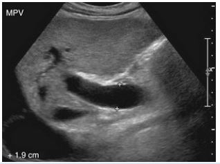
Figure 1: USG showing measurement of Portal vein diameter.
Ultrasound is a valuable tool for diagnosing and managing liver disease, providing accurate and reliable information that can guide treatment decisions and improve patient outcomes [8]. In this study the mean portal vein diameter was 8.33±1.78 mm. similar findings have been reported in other studies conducted in Nepal: Regmi S et al [1], found 9.01±1.24 mm and Jha AK et al [8], found 9.57±0.66 mm. The consistency in the reported portal vein diameter measurement across different studies and populations may be attributed to the use of similar methodologies, such as the trans-abdominal approach and comparable probe frequencies.
Studies conducted in various countries have reported similar findings regarding the mean portal vein diameter measured by ultrasound. S. Albagir et al [2], reported a mean value of 10.4±1.22 mm among Arabians, while Saha et al [7], reported a mean value of 8.81 mm among Bangladesh. Similarly, Bhattacharya et al [3], in West Bengal, India, reported a mean value of 10.12±0.89 mm. Numerous studies have been conducted on the measurement of the portal vein diameter using ultrasound have shown consistent values across different countries, ethnic groups, and races, with minimal variation. This suggests that the technique is reliable and can be applied uniformly across diverse populations.
In this study, the mean portal vein diameter was found to be slightly higher in males than in females, statistically significant at p<0.05 level. This finding is consistent with the study by S. Albagir et al [2] but contrasts with the study by Jha AK et al [8]. The mean portal vein diameter across different age group was highest in individuals aged 20 years and below, with a mean diameter of 8.91±1.69 mm and lowest in age group 21-30 years with mean portal vein diameter of 7.73±1.53 mm; however, this was not statistically significant (p>0.05).
This study found no significant correlation (p>0.05) between portal vein diameter and age, consistent with the results of other studies such as S. Albagir et al [2], Gareballah et al [6] who also reported no significant influence of age on portal vein diameter. Various studies have investigated the effect of height on portal vein diameter, yielding mixed results. In this study, a positive correlation was observed between height and portal vein diameter (p<0.05). This finding aligns with the results of other studies, including Gareballah et al [6], and Jha Ak et al [8], which also identified a positive correlation between height and portal vein diameter. These findings suggest that portal vein diameter increases with height, likely due to fundamental anatomical and physiological scaling.
Studies examining the relationship between portal vein diameter and BMI have reported mixed findings. Bhattacharya et al [3] found a positive correlation between BMI and portal vein diameter in individuals with chronic liver disease. Similarly, studies by Saha et al [7] and Jha AK et al [8] have also reported a positive correlation. However, Gareeballah et al [6] did not find a statistically significant influence of BMI on portal vein diameter. This study showed no significant correlation between PV diameter and BMI (p>0.05).
This hospital-based descriptive cross-sectional study of 193 apparently healthy adults established the normal portal vein diameter in healthy Nepalese adults to be 8.33±1.78 mm. The mean portal vein diameter in males was slightly higher than in females which may be attributed to the higher number of female participants. There was a significant positive correlation between portal vein diameter and both gender and height suggesting that these factors, along with age and other anthropometric variables, may influence portal vein diameter.
Funding
This study was not supported by any funding.
Conflict of interest
The authors declare that they have no conflict of interest
We always work towards offering the best to you. For any queries, please feel free to get in touch with us. Also you may post your valuable feedback after reading our journals, ebooks and after visiting our conferences.

